Describe the change
Water-soaked, soft or disintegrating tissue.
 MushroomFarming.in
A joint educational initiative by

 ICAR–DMRSolan
Bacterial diseases
Wet, collapsing tissue is a reason to separate produce and investigate timing. It can be a primary infection or secondary decay after injury.
Organism / cause: Several organisms; Burkholderia gladioli pv. agaricicola is one researched example
Where this evidence applies: Symptom differential. The cited Burkholderia evidence includes Agaricus bitorquis; it is not the cause of every soft rot on this crop list.
Picture → observation → next check
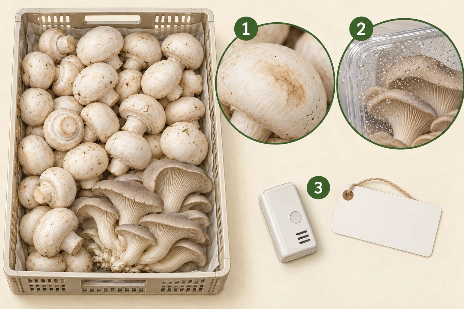
Ask whether it appeared before picking, during handling or in storage. A brown area is not automatically bacterial blotch.
Record condensation, temperature changes and elapsed time. Do not use a visual match to release a suspect lot.
Link harvest, packing and transport records so affected produce can be traced and assessed.
Water-soaked, soft or disintegrating tissue.
Heat or chilling injury followed by secondary decay.
Reconstruct the harvest-to-storage timeline.
A visual match is only a starting point. A laboratory must identify the organism and interpret whether it caused the primary damage.
01 · Observe
No verified reusable diagnostic photograph is included for this entry. The illustration above explains observation context, not a confirmed disease specimen. Consult the original source figures where available.
02 · Compare
Damaged wet tissue and contaminated equipment can sustain contamination; the initial cause may be earlier than visible collapse.
03 · Check
What confirmation needs: A laboratory must identify the organism and interpret whether it caused the primary damage.
Use the field record and sampling checklist →04 · Respond
These are investigation and containment steps, not a guaranteed cure. Do not eat, sell or distribute suspect material on the strength of a visual match. Do not culture unknown contaminants or experiment with pesticides in a growing room.
05 · Learn
After intervention, compare new affected units, crop condition and inspection results against the same recorded baseline. A lack of visible growth does not, by itself, verify decontamination or food safety.
Read the stage-by-stage prevention plan →Research examples support a differential, not a universal pathogen assignment.
Research review, PMC · 2008
Used for: Bacterial soft rot and yellowing; Burkholderia gladioli pv. agaricicola in Agaricus bitorquis.
Limit: Research taxonomy and mechanism, not evidence that all soft rot is this organism.
Research article, PMC · 2020
Used for: P. agarici gill disease and P. tolaasii blotch distinctions.
Limit: Experimental phage results are not a farm treatment protocol.
Food & worker safety
Separate when damage started from when someone noticed it. That distinction prevents a storage problem being mislabelled as a growing-room disease.
Harvest, packing, transport and storage; a crop can deteriorate after leaving a healthy room.
Signs, checks & response →Food & worker safety
A healthy-looking mushroom is not proof of food safety. A crop-disease photo guide cannot detect Listeria, Salmonella or unacceptable residues.
All edible-mushroom food chains; enoki outbreak evidence is a specific example, not a claim about every crop.
Signs, checks & response →Environmental disorders
A room-air reading can miss a hot substrate core. Equally, a cold chain suitable for button mushrooms must not be assumed suitable for paddy straw.
Substrate, crop and storage temperatures must be interpreted separately for each species.
Signs, checks & response →