Describe the change
Yellow, ginger or brown lesions on the cap.
 MushroomFarming.in
A joint educational initiative by

 ICAR–DMRSolan
Bacterial diseases
“Ginger blotch” and “yellow blotch” describe appearances, not a dependable field identification of one bacterium.
Organism / cause: Multiple Pseudomonas lineages; species cannot be read from cap colour
Where this evidence applies: Blotch-associated isolates from Agaricus in Western European research.
Picture → observation → next check
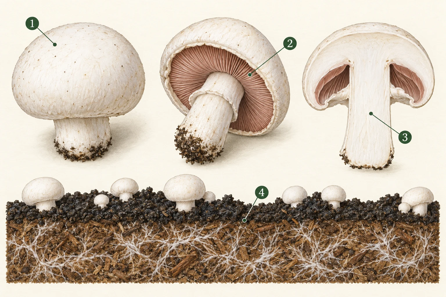
Record a local spot versus whole-cap colour, and whether it was present before picking.
Separate normal maturity-related pigmentation from unusual wetness, exudate or deterioration.
Internal changes can differ from the outside. Obtain sampling instructions before cutting suspect material.
Note whether the change starts in the fruit body, the casing surface or the substrate below.
Yellow, ginger or brown lesions on the cap.
P. tolaasii blotch, mechanical injury and residues.
Use neutral-light photographs and record picking/packing timing.
A visual match is only a starting point. Molecular characterisation and disease testing underpin the published classifications; a visual match is weaker evidence.
01 · Observe
No verified reusable diagnostic photograph is included for this entry. The illustration above explains observation context, not a confirmed disease specimen. Consult the original source figures where available.
02 · Compare
Wet surfaces and handling are investigation priorities; different isolates can behave differently.
03 · Check
What confirmation needs: Molecular characterisation and disease testing underpin the published classifications; a visual match is weaker evidence.
Use the field record and sampling checklist →04 · Respond
These are investigation and containment steps, not a guaranteed cure. Do not eat, sell or distribute suspect material on the strength of a visual match. Do not culture unknown contaminants or experiment with pesticides in a growing room.
05 · Learn
After intervention, compare new affected units, crop condition and inspection results against the same recorded baseline. A lack of visible growth does not, by itself, verify decontamination or food safety.
Read the stage-by-stage prevention plan →2020 molecular research; not an India-specific prevalence survey.
Research article indexed in PubMed · 2020
Used for: Blotch can involve several Pseudomonas species; molecular and pathogenicity testing.
Limit: Abstract and figure captions reviewed. Cap colour cannot distinguish the organisms.
Research article, PMC · 2020
Used for: P. agarici gill disease and P. tolaasii blotch distinctions.
Limit: Experimental phage results are not a farm treatment protocol.
Bacterial diseases
Persistent surface wetness can favour bacterial blotch. Managing cap drying means balancing the crop environment, not simply drying the entire room.
Agaricus caps; a brown spot does not establish bacterial identity.
Signs, checks & response →Bacterial diseases
The gill surface matters: some disease is hidden beneath the cap. A cap-only photograph can miss the most useful evidence.
Agaricus bacterial gill disease.
Signs, checks & response →Environmental disorders
Unexpected forms or lesions after cleaning, spraying or nearby fuel use need an exposure history as well as a disease differential.
Rosecomb is classically described in Agaricus; broader crop filters cover exposure investigation, not identical symptoms.
Signs, checks & response →