Describe the change
Discoloured or deteriorating gills with droplets or exudate.
 MushroomFarming.in
A joint educational initiative by

 ICAR–DMRSolan
Bacterial diseases
The gill surface matters: some disease is hidden beneath the cap. A cap-only photograph can miss the most useful evidence.
Organism / cause: Pseudomonas agarici
Where this evidence applies: Agaricus bacterial gill disease.
Picture → observation → next check
Record a local spot versus whole-cap colour, and whether it was present before picking.
Separate normal maturity-related pigmentation from unusual wetness, exudate or deterioration.
Internal changes can differ from the outside. Obtain sampling instructions before cutting suspect material.
Note whether the change starts in the fruit body, the casing surface or the substrate below.
Discoloured or deteriorating gills with droplets or exudate.
Water droplets after handling and general tissue decay.
Photograph cap, underside and a comparison mushroom from the same lot.
A visual match is only a starting point. Bacterial isolation and identification distinguish P. agarici from other causes of gill breakdown.
01 · Observe
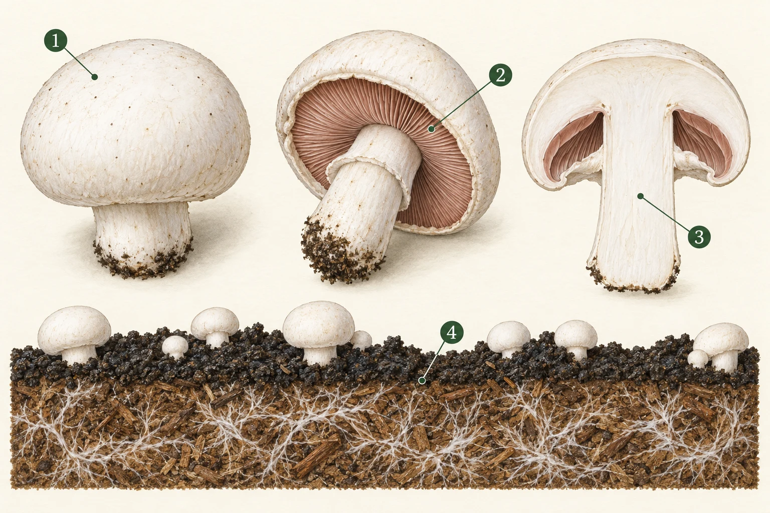
No verified reusable diagnostic photograph is included for this entry. The illustration above explains observation context, not a confirmed disease specimen. Consult the original source figures where available.
02 · Compare
Bacterial contamination and wet handling conditions should be traced; the precise source needs local investigation.
03 · Check
What confirmation needs: Bacterial isolation and identification distinguish P. agarici from other causes of gill breakdown.
Use the field record and sampling checklist →04 · Respond
These are investigation and containment steps, not a guaranteed cure. Do not eat, sell or distribute suspect material on the strength of a visual match. Do not culture unknown contaminants or experiment with pesticides in a growing room.
05 · Learn
After intervention, compare new affected units, crop condition and inspection results against the same recorded baseline. A lack of visible growth does not, by itself, verify decontamination or food safety.
Read the stage-by-stage prevention plan →Research on mushroom-pathogenic pseudomonads; phage experiments are not an available cure.
Research article, PMC · 2020
Used for: P. agarici gill disease and P. tolaasii blotch distinctions.
Limit: Experimental phage results are not a farm treatment protocol.
Bacterial diseases
Persistent surface wetness can favour bacterial blotch. Managing cap drying means balancing the crop environment, not simply drying the entire room.
Agaricus caps; a brown spot does not establish bacterial identity.
Signs, checks & response →Bacterial diseases
Wet, collapsing tissue is a reason to separate produce and investigate timing. It can be a primary infection or secondary decay after injury.
Symptom differential. The cited Burkholderia evidence includes Agaricus bitorquis; it is not the cause of every soft rot on this crop list.
Signs, checks & response →Food & worker safety
Separate when damage started from when someone noticed it. That distinction prevents a storage problem being mislabelled as a growing-room disease.
Harvest, packing, transport and storage; a crop can deteriorate after leaving a healthy room.
Signs, checks & response →