


Wet Bubble Disease
An evidence-led guide to recognise Mycogone-associated patterns while differentiating dry bubble and soft injury.
See the symptom. Check the cause. Act carefully.
Real research photographs with practical, nonchemical first steps. These examples come from published studies, not MushroomWale farms. Resemblance is a clue, not confirmation of disease in your crop.
- 01Observe the crop and record the pattern
- 02Limit transfer to clean areas
- 03Check the diagnosis and follow a response plan
- 04Review the next crop for recurrence
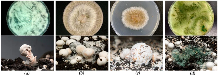
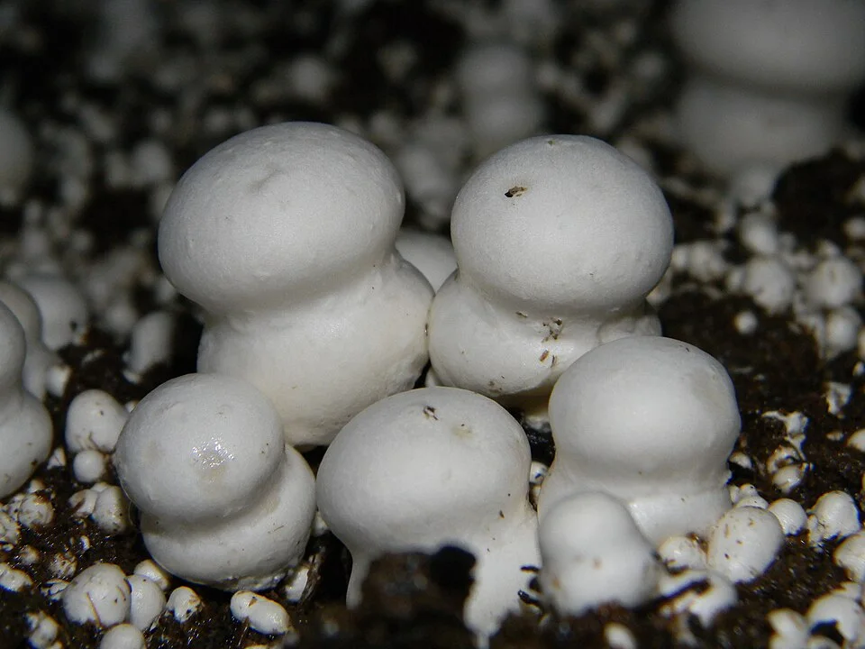
Wet bubble
Irregular swollen tissue, severely distorted mushrooms and amber droplets are useful clues. Decay can develop as affected tissue deteriorates.
- What to do first
- Mark the area and avoid splashing it. Once wet bubble is identified, trained staff can carefully enclose local affected material in a disposable bag, seal it and follow the farm's containment and disposal procedure.
- Reduce the chance of recurrence
- Protect casing and water supplies from soil, dust and crop debris. Keep clean equipment away from contaminated floors and spent material.
- What can look similar?
- Do not apply this removal approach to an unidentified bubble: dry bubble handling differs. Ask a crop-health specialist to distinguish them.
- What to record
- Record the casing lot, first affected location, water source, spread after watering and photographs before intervention.
From symptom to cause
A first look, not a diagnosis. Source: ICAR-DMR manual and disease bulletin.
| What you see | Likely cause | First step |
|---|---|---|
| Long stems, small caps | CO₂ too high (above about 1,500 ppm) | More fresh air |
| Green patches on compost | Trichoderma; low pH or leftover ammonia | Isolate, remove; check next batch |
| No pins after casing | Room not aired and cooled | 16 to 18 °C, ventilate |
| Brown sunken spots on caps | Bacterial blotch; wet caps | Dry the surface, humidity under 85% |
| White wet lumps | Wet bubble from casing | Salt, remove; steam next casing |
| Small flies | Sciarid or phorid flies | Screens, light traps, hygiene |
Keep clean and suspect work separate
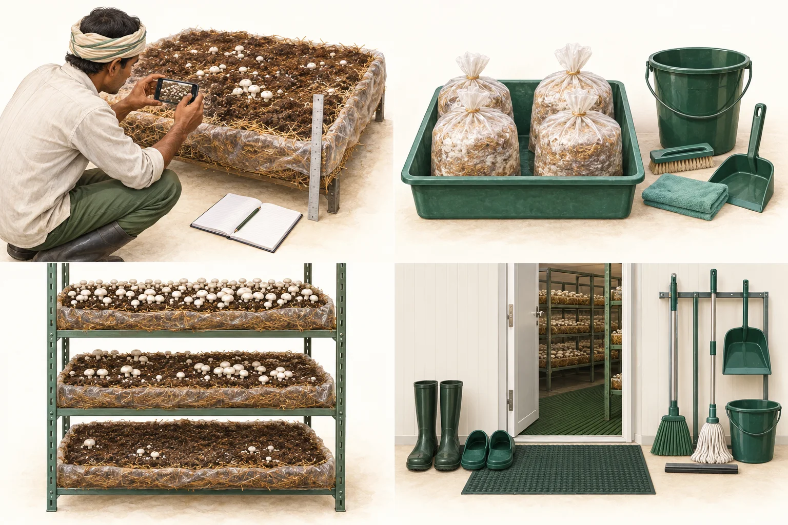
AI-generated educational illustrations. Not actual farm photographs, diagnostic evidence or construction specifications.
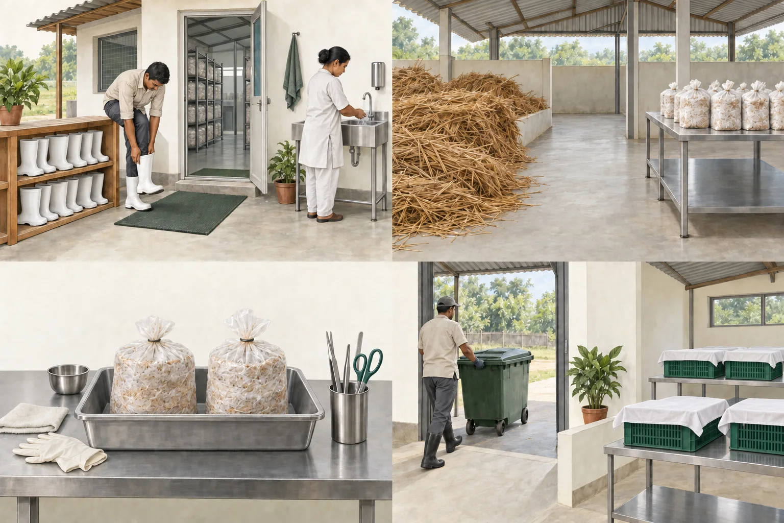
Make clean entry a routine
Clean entry has a place and a repeatable sequence. Footwear, hands and movement between areas are part of farm hygiene. The illustration does not demonstrate a validated disinfection process.
Assign the entry routine, provide suitable facilities and check that visitors and workers follow it.
Protect prepared material
Raw inputs and protected prepared material are separated. Treatment does not protect a substrate indefinitely. Handling afterwards can introduce contamination.
Keep work areas, tools and movement routes separated. Label batches so a later problem can be traced to its preparation history.
Keep suspect bags closed
A separate tray helps identify material being held for investigation. The bags are illustrative and not diagnosed. Opening, shaking or smelling suspect material is not a confirmation method.
Mark the batch, limit movement and seek qualified advice on sampling or disposal. Do not return dedicated tools to clean work without the appropriate procedure.
Plan the spent-material route
Used material and saleable harvest follow different handling paths. A disposal route can undo clean-work separation if it crosses incoming materials or harvest handling.
Map the exit route and cleaning responsibility. Check the actual workflow during a busy room turnaround, not only on a drawing.
Wet bubble
Current causal description: Hypomyces perniciosus (syn. Mycogone perniciosa)
Symptom progression
Timing of infection can change what appears.
Abnormal undifferentiated tissue
White mouldy growth on developing mushrooms
Large irregular swollen masses
Malformed mushrooms with swollen stipes or deformed caps
Wart-like growths or cap spotting
Numerous amber to golden-brown exudate droplets
Softening and putrefaction
Foul odour may develop
Could it be something else?
Retain alternatives until discriminating evidence is present.
Patterns to map
- Can appear as isolated affected pieces before wider secondary spread
- Water, handling and vectors may move contamination
Confused with
- Dry bubble
- Bacterial soft rot
- Severe mechanical injury
How to confirm
- Compare droplet number, tissue wetness and progression with dry bubble
- Microscopy or culture by a qualified laboratory
- Specialist inspection where distribution is expanding
Integrated management hierarchy
Prevention and verified root-cause correction come before chemical action.
- 1
Do not touch or spread suspect tissue
- 2
Document and restrict movement
- 3
Inspect the surrounding area
- 4
Escalate rapid or repeated outbreaks
- 5
Treat historic chemical references as non-operational
Follow the chain upstream
A name is not a root cause.
- Visible crop change
- Map the pattern
- Check process and environment
- Verify the mechanism
- Correct the root cause
Quick recognition
Use wet bubble disease to recognise Mycogone-associated patterns while differentiating dry bubble and soft injury. Recognition begins the investigation; it does not confirm a cause.
- Observe early, developing and advanced signs
- Map distribution and progression
- Compare biological and abiotic causes
- Confirm before high-risk action
What to record
Capture crop stage, crop day, room, rack, layer, pattern, first observation, progression, compost and casing batches, watering, chemistry, pests and timestamped environmental readings.
Could it be something else?
Compare visual appearance with spatial pattern, progression, environmental history, process history and missing expected signs. Colour or one photograph is weak evidence.
Evidence still needed
State the unanswered questions and next measurement. Useful evidence can be visual, environmental, spatial, temporal, microscopic, laboratory, molecular, trap-based or process-history based.
Immediate safe direction
Document the affected area, avoid unnecessary disturbance or cross-room movement, isolate suspect tools, inspect adjacent areas and preserve representative evidence. Do not spray automatically.
Root-cause review
Trace the visible problem through water, evaporation, airflow, sensor placement, compost, casing, staff movement, flies, tools or room design where evidence supports the connection.
Chemical and regulatory boundary
Historical management literature is separated from current practice. No crop-protection dose is published unless the exact product, formulation, mushroom crop label, target, method, harvest interval, worker-safety information and current Indian status are verified.
When remote guidance is inadequate
Seek technical inspection or laboratory support for rapid unexplained loss, repeated outbreaks, suspected virus, unknown mould, severe pests, chemical injury, multiple affected rooms or unresolved contradictions.
Trace the claim and its limits.
Pathogen biology, dated advisories and current regulatory status are stored separately.
Diseases and Competitor Moulds of Mushrooms and their Management
Indian mushroom disease descriptions, symptoms, epidemiology, historical taxonomy and competitor-mould records.
Open sourceICAR-DMR Technical Bulletin Library
Institutional publication index for disease, farm-design, post-harvest and spent-substrate references.
Open sourceFungal diseases of mushrooms and their control
Dry bubble, wet bubble and cobweb symptom progression and hygiene pathways.
Open sourceDirectorate of Plant Protection, Quarantine and Storage regulatory functions
Indian pesticide registration and regulatory-governance context under the Insecticides Act and Rules.
Open sourceChemical governance is a publication gate.
No operational pesticide, fungicide, insecticide, disinfectant or fumigant dose is published. Historical references are not current labels.
- Current operational dose
- Not published
- Mushroom crop label
- Not established by this record
- Review status
- Regulatory review required