Describe the change
A separate white colony becoming green in substrate.
 MushroomFarming.in
A joint educational initiative by

 ICAR–DMRSolan
Fungal diseases
A bag can contain competing mould before green colour is visible. Oyster-associated Trichoderma species differ from the main button-mushroom epidemic species.
Organism / cause: Trichoderma species; oyster-associated T. pleuroti and T. pleuroticola
Where this evidence applies: Detailed pathogen evidence exists for Pleurotus; the other listed crops have published green-mould host reports, not a shared diagnosis.
Picture → observation → next check
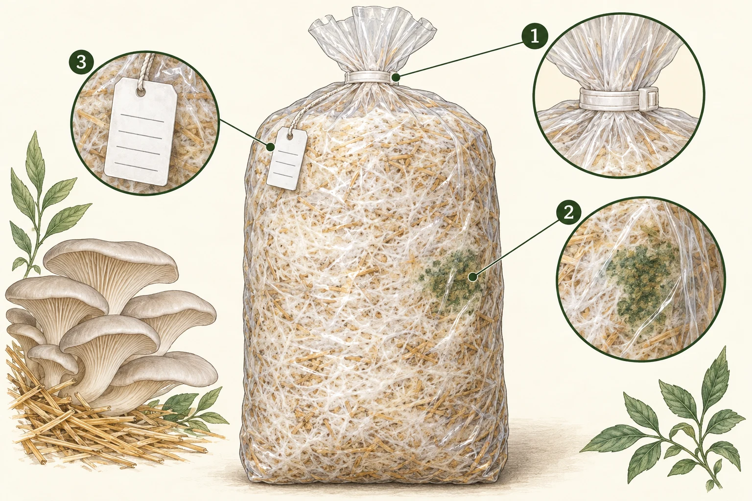
Record whether the first change is near a seal, filter, tear or fruiting cut. Do not open the bag to investigate.
Photograph its position and change over time. A green patch does not identify a genus or species; early growth may be white.
Keep spawn lot, substrate treatment, inoculation date and rack position together. Compare other bags from the same batch.
A separate white colony becoming green in substrate.
Other green-sporing fungi.
Record whether the patch starts at the inoculation point, seal, cut or throughout the bag.
A visual match is only a starting point. A laboratory can distinguish Trichoderma taxa; colour and host name are insufficient.
01 · Observe
No verified reusable diagnostic photograph is included for this entry. The illustration above explains observation context, not a confirmed disease specimen. Consult the original source figures where available.
02 · Compare
Introduction can occur before or after substrate treatment; spawn handling, cooling, bag closure and later handling need separate examination.
03 · Check
What confirmation needs: A laboratory can distinguish Trichoderma taxa; colour and host name are insufficient.
Use the field record and sampling checklist →04 · Respond
These are investigation and containment steps, not a guaranteed cure. Do not eat, sell or distribute suspect material on the strength of a visual match. Do not culture unknown contaminants or experiment with pesticides in a growing room.
05 · Learn
After intervention, compare new affected units, crop condition and inspection results against the same recorded baseline. A lack of visible growth does not, by itself, verify decontamination or food safety.
Read the stage-by-stage prevention plan →Molecular research and detection review; host records do not imply equal incidence across crops.
Komon-Zelazowska et al., Applied and Environmental Microbiology · 2007
Used for: Molecular distinction of oyster-associated green-mould taxa from the Agaricus epidemic organism.
Limit: Historical nomenclature includes T. pleurotum; current papers commonly use T. pleuroti. A green patch is not species confirmation.
Biology · 2023
Used for: Detection limits and reported hosts including milky, paddy straw and Ganoderma.
Limit: Host reports do not show prevalence in every Indian region; molecular identification needs qualified interpretation.
Mycobiology · 2013
Used for: Bottle contamination, competing fungal growth and failure of primordia.
Limit: Selected Korean farms; association and identification are not universal proof of causation for every isolate.
Fungal diseases
Green mould may damage block colonisation and fruiting. It must not be confused with the normal development of a brown, mature shiitake block surface.
Lentinula edodes sawdust blocks; log production has a different contamination environment.
Signs, checks & response →Competitor moulds
Green, blue-green or dark growth is not automatically Trichoderma. Neither harmlessness nor toxin production can be established from its colour.
Raw-material, spawn, substrate and storage contamination differential across production systems.
Signs, checks & response →Viruses & culture health
Separate three questions: was the spawn viable, was the substrate suitable, and was the inoculation/handling process sound?
Applies from grain spawn to compost, straw and supplemented wood substrates.
Signs, checks & response →